External Laser Therapy
The Principle
In external therapies, lasers are successfully used in pain reduction, rehabilitation, stroke treatment, acupuncture, dermatology and cosmetics, dentistry and veterinary medicine.


How does it work?
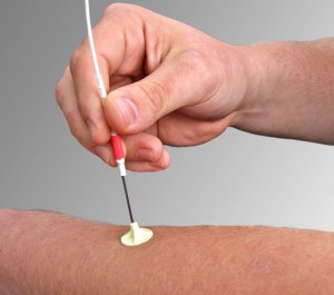
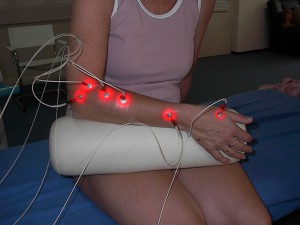
Red, infrared, green and blue lasers are used for different penetration depths and for different biological effects in the tissue. The lasers can be fixed to the skin with self-adhesive tubes or put together in special applicators for radiation of bigger areas. The highly focused laserlight enters the tissue and leads to biostimulating and reparative effects on the tissue without any side-effects.
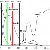
The picture shows the absorption spectrum of our skin which leads to the different penetration depths of the different lasers.
How can these effects be explained?
- laserlight leads to a chain of biochemical reactions through specific absorption (cytochromes, porphyries)
- The pigments of the respiratory chain are particularly suited for the specific absorption of irradiation to trigger photochemical reactions
- Light electrons are stimulated in the respiratory redux chain
- The electrons are transported against the redux decline in the respiratory chain, which finally leads to a phosphorylization from ADP to ATP and to increased membrane potential
- An increase of the ATP production of 150% can be evidenced through infrared exposure of
yeast. - The skin poorly resorbs irradiation in the near infrared range, in particular between 800 and
900 nm, which enables the irradiation to penetrate the tissue relatively deep - For medical treatment, red light in a visible range (630 – 680 nm) and infrared light in a range between 800 and 900 nm with a significantly higher penetration depth is used almost exclusively
What are the physiological effects of laser light?
- Proliferation on immune cells leads to the combat of inflammations and an accelerated healing of wound as well as an increased endorphin disbursement, increase of the ATP production and increased nervous cell potential
- Increased leukocyte phagocytosis, boosted neovascularisation, increased collagen
formation and protein biosynthesis. It also leads to an improved cell respiration and
stabilisation of the membrane potential - Enhancement of the proton gradient via the mitochondria membrane, generation of an increased potential difference with increased phosphorisation of ATP (increase of 150%)
- No modification of intact cells
- Energetic build-up of sick cells
- Energy is to a large extent (more than 40%) used for ATP synthesis, in order to increase the pump activity for maintaining the membrane potential
- Membrane stabilisation leads to blocking of impulses, reducing the transmissions of pain sensations
- The cell’s calcium content is regulated (diminished ATP synthesis leads to a an overflow of the cell with calcium and activation of proteinasis, resulting in the death of the cell, the necrosis)
- In the pre-necrotic state, cells suffer from acute lack of energy with sodium and calcium streaming in, which can only be removed with the utmost pump activity. This pump activity can only be enhanced by radiant energy


